
If you've been told you need a stent, you may picture a procedure that hasn't changed in years. In fact, coronary angioplasty has advanced considerably over the past decade — becoming more precise and, in selected patients, safer.
The short answer
Angioplasty has advanced considerably over the past decade:
- Imaging from inside the artery (IVUS and OCT) guides stent size and expansion.
- Drug-coated balloons can treat selected blockages without leaving metal behind.
- Lithotripsy and atherectomy prepare heavily calcified arteries so stents can open fully.
- Thinner modern stents mean many patients need a shorter course of dual blood thinners.
Four Advances That Are Changing Angioplasty
The stent remains central to treatment, but how a blockage is assessed, prepared, and treated has transformed. Here are the four most important advances — explained in plain language for patients and referring GPs.
1. Imaging Inside the Artery
IVUS and OCT probes reveal what an angiogram cannot — wall thickness, calcium, and stent expansion.
2. Drug-Coated Balloons
Treat selected blockages with medication and no permanent metal implant left behind.
3. Calcium-Modification Tools
Lithotripsy and atherectomy crack or sand down hardened plaque so stents can open fully.
4. Modern Stents & Shorter DAPT
Thinner-strut stents heal faster, and many patients now need fewer months of dual blood-thinning tablets.
1. Looking Inside the Artery: IVUS and OCT

OCT: the artery wall in fine detail, each stent strut a bright dot against the wall.
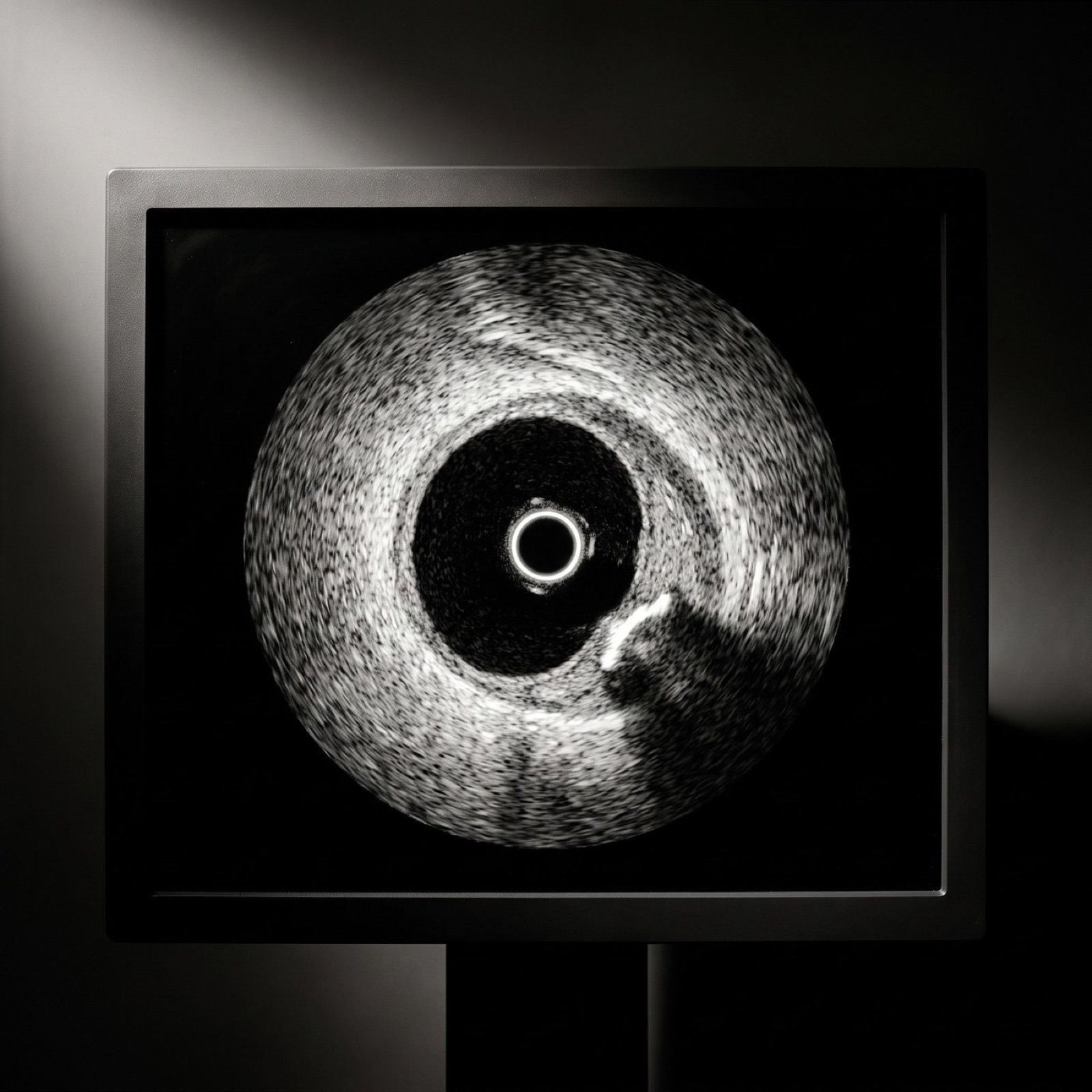
IVUS: plaque thickens one side; calcium is a bright arc with a dark shadow behind it.
Intravascular ultrasound (IVUS) and optical coherence tomography (OCT) are miniature imaging probes — about the width of a few hairs — passed into the artery on a thin wire. They show the vessel from the inside, in cross-section, rather than relying only on the dye-based X-ray outline (the angiogram).
An angiogram shows the shape of the channel, but not the thickness of the artery wall, how much calcium is present, or whether a stent has opened fully. Imaging from inside answers all three — helping choose the right stent size, land it in healthy segments, and confirm it is fully expanded and pressed against the wall.
IVUS & OCT: What the Evidence Shows
Several large trials now support imaging guidance for complex blockages, and the results are compelling:
RENOVATE-COMPLEX-PCI (NEJM, 2023)
1,639 patients. Imaging guidance cut the combined risk of cardiac death, heart attack, or repeat procedure from 12.3% to 7.7% at a median of 2.1 years — and the benefit persisted with longer follow-up.
OCTOBER (NEJM, 2023)
1,201 patients with bifurcation lesions. OCT guidance reduced major adverse cardiac events at two years: 10.1% vs 14.1% with angiography alone.
ILUMIEN IV (NEJM, 2023)
2,487 patients. OCT guidance produced larger, better-expanded stents and fewer stent clots, although the overall rate of repeat problems in the treated artery was not significantly lower at two years.
On this evidence, the 2024 ESC guideline gives intracoronary imaging a Class I recommendation for guiding stenting in anatomically complex lesions — particularly left main, true bifurcation, and long lesions. Trials suggest imaging guidance can reduce repeat procedures and stent-related problems in the years that follow.
2. Treating Without a Permanent Stent: Drug-Coated Balloons
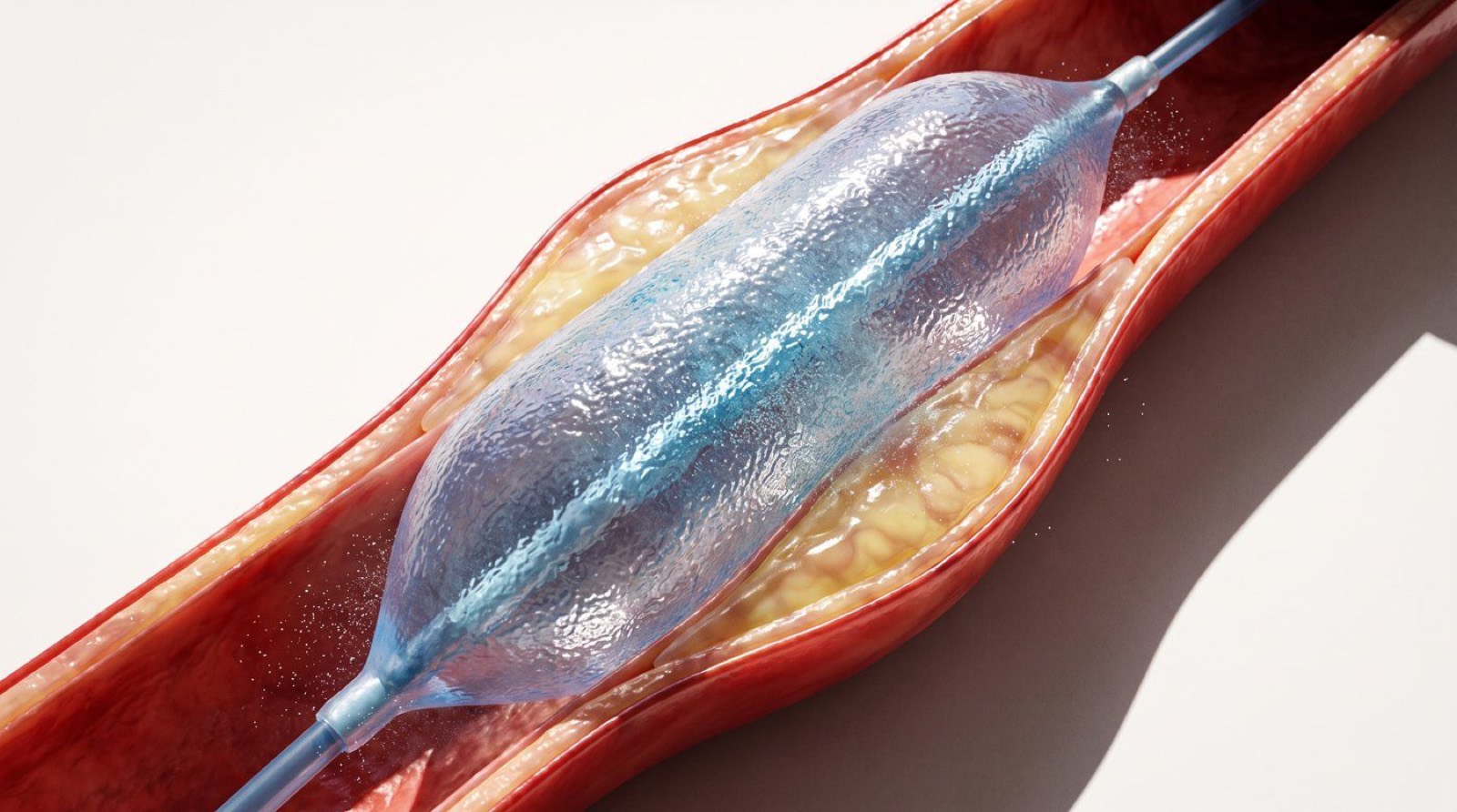
A drug-coated balloon is an angioplasty balloon whose surface carries a medication — usually paclitaxel or sirolimus. During a brief inflation of 30–60 seconds, the drug is pressed into the artery wall. The balloon opens the narrowing and leaves the drug behind to discourage re-narrowing, but no metal stays in the artery.
Avoiding a permanent implant is attractive in certain situations: inside a stent that has re-narrowed, in very small arteries where a stent would leave little room, and in people who cannot take long courses of blood-thinning tablets.
| Setting | Key Trial Finding | Current Position |
|---|---|---|
| Re-narrowed stent (in-stent restenosis) | AGENT IDE: fewer target-lesion failures at 1 year (17.9% vs 28.6%) | Established option |
| Small native arteries (<3 mm) | BASKET-SMALL 2: comparable to drug-eluting stents at 3 years | Reasonable option |
| Larger native arteries | REC-CAGEFREE I: balloon-first not as good as up-front stent at 2 years | Stents remain preferred |

3. Hard, Calcified Arteries: Lithotripsy and Atherectomy
Over years, some plaque hardens into calcium that can be as stiff as bone and may form a complete ring inside the artery. Calcium-modification tools prepare these arteries before a stent goes in — because calcium stops a stent from opening fully, and an under-expanded stent is the one most likely to fail.
Intravascular Lithotripsy (IVL)
A balloon emits sonic pressure waves to crack calcium from within — similar in principle to shockwave treatment for kidney stones. In Disrupt CAD III, 92% of patients were free of major cardiac events at 30 days with good stent expansion.
Rotational & Orbital Atherectomy
A tiny, fast-spinning, diamond-coated tip sands down hardened plaque. These are long-established, guideline-supported tools. Note: the ECLIPSE trial (Lancet, 2025) found routine orbital atherectomy did not improve one-year outcomes over careful balloon preparation in lesions suitable for either approach.
These techniques have made some heavily calcified arteries treatable by keyhole angioplasty when, in the past, bypass surgery may have been the only realistic option. Which tool is used depends on the pattern and depth of calcium — exactly what intracoronary imaging reveals.
4. Modern Stents and Shorter Blood-Thinning Courses
Thinner-Strut Stents
Today's drug-eluting stents have struts roughly half the thickness of earlier generations, with improved coatings and drugs. Thinner struts heal over faster and are less likely to trigger clots or re-narrowing.
Shorter Dual Antiplatelet Therapy (DAPT)
Partly because of this, there has been a clear shift towards shorter courses of two blood-thinning tablets (usually aspirin plus a second agent) in suitable patients. Trials show that shortening dual therapy to 1–3 months and then continuing one tablet reduces bleeding without a clear rise in heart attack or stent clots. After a stent for stable angina the usual default is six months (shorter if bleeding risk is high). After a heart attack it is usually 12 months, sometimes shortened by your cardiologist.
Important
The right duration is individualised. Never stop blood-thinning tablets without speaking to your cardiologist first — even if a relative with a stent took them for a different length of time.

What Has Not Changed: The Foundation of Heart Care
For stable coronary artery disease, medication and risk-factor control remain the foundation of treatment. The landmark ISCHEMIA trial (Maron et al., NEJM, 2020) showed that in stable patients, an early procedure-based strategy did not reduce death or heart attack overall compared with good medical therapy — although it relieved angina more effectively in people with frequent symptoms.
The advances described in this article improve how a procedure is done — more precise, safer, and longer-lasting. They do not change who needs one.
Control Blood Pressure
Consistently managed blood pressure reduces strain on coronary arteries and lowers event risk.
Manage Cholesterol & Diabetes
Lipid-lowering and glucose control slow plaque progression with or without a stent.
Don't Smoke
Smoking cessation is one of the most powerful interventions available to any cardiac patient.
Stay Active
Regular physical activity and taking prescribed medicines remain essential alongside any procedure.
For Referring GPs: When to Consider Complex or Imaging-Guided PCI
Consider referral to a centre experienced in imaging-guided and complex angioplasty for patients with any of the following. The 2024 ESC guideline gives a Class I recommendation for intracoronary imaging to guide stenting in left main and anatomically complex lesions.
Heavily Calcified Coronary Arteries
Identified on CT or prior angiography — calcium-modification tools may be required.
Left Main or Bifurcation Disease
Anatomically complex lesions where imaging guidance is now a Class I recommendation.
Chronic Total Occlusion
A completely blocked artery with ongoing symptoms — specialist techniques required.
Prior Stent Failure
In-stent restenosis or stent thrombosis — drug-coated balloons and imaging guidance are key.
Multivessel Disease
Where the choice between stenting and bypass surgery is unclear and requires careful assessment.
Questions to Ask Your Cardiologist Before a Procedure
Being informed helps you get the best outcome. Before your angioplasty, consider asking your cardiologist these five key questions:
Will you use imaging from inside the artery?
Ask whether IVUS or OCT will guide your stent placement.
Is my artery heavily calcified?
And if so, how will it be prepared before the stent goes in?
Is a stent the best option for me?
Or could a drug-coated balloon be appropriate for my blockage?
How long will I need two blood-thinning tablets?
And what is the reason for that specific duration in my case?
What can I do with medication and lifestyle?
To get the best possible long-term result from my procedure.
For GPs
- Referral triggers for complex PCI review: left main or true bifurcation disease, heavy calcification, in-stent restenosis or stent thrombosis, symptomatic CTO, or multivessel disease where PCI versus CABG is unclear.
- DAPT defaults in letters: about 6 months after elective PCI and about 12 months after ACS; 1–3 months if bleeding risk is high. The letter should state the stop date. (ESC 2024 CCS; ESC 2023 ACS)
- Don't stop the P2Y12 inhibitor early for dental work, minor skin surgery or colonoscopy without polypectomy. Defer elective non-cardiac surgery ideally 6 months after elective PCI and 12 months after ACS; if it can't wait, discuss with the cardiologist (aspirin usually continued).
- PPI while on DAPT, especially over 65 or with prior GI bleed; avoid NSAIDs. Ticagrelor breathlessness is usually benign: review rather than stop.
- Long term: single antiplatelet (aspirin or clopidogrel) lifelong unless on an anticoagulant.
General information for referring practitioners. Check current MBS and PBS criteria before acting.